


The causes of thoracic vertebrae degeneration are not fully elucidated.The most important are the hereditary tendencies and age-related changes in the disc.
Osteocartilage in the chest and spine: Symptoms.
The first stage of neurological complications of osteochondrosis in the chest spine.
Clinical manifestations are related to reflex muscle tone.Dorxago (Breast Background).Acute pain in the chest area associated with exercise.The intensification suddenly began.The amount of exercise of the thoracic spine is strictly limited."Stone" density of secondary muscles.Dorsago's proliferation should not exceed 7-10 days after proper treatment.
Back pain (back pain).PACENERS complains about moderate pain in the chest area, which is exacerbated during exercise, during exercise or at a certain position.The beginning is usually gradual.Clinically, the curvature of spine, tension and soreness is often determined.In most cases, pain takes 2-3 weeks, but in the absence of treatment, a chronic course can be performed.
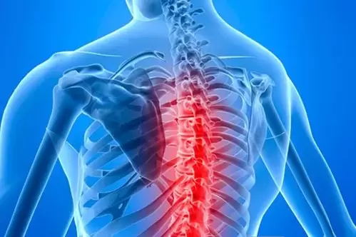
Fruit flies (chest pain).Breast pain is one of the most common complaints patients have when they visit their doctor.In this case, the differential diagnosis is performed by heart disease (angina, myocardial infarction).
Chest pain usually occurs in the context of osteochondrosis in the spine of the chest.The pain is deep, broken, sore, aggravated, exercise or staying in one position for a long time.In the chest area, limited exercise, tension and pain, on palpation of the side muscles.
Syndrome of the chest wall, the stupidity, soreness of the front surface of the chest, prolonged pain occurs during manual exercise and turns.Taking nitroglycerin under the tongue will not stop the pain.Weird spots were found in the chest muscles of the size.
Stage 2 of neurological complications of osteochondrosis in the chest spine.
Hernia of the thoracic disc is extremely rare with radiation syndrome.This is due to the structure of the thoracic vertebrae.Compression of the spine (or intercostal neuralgia) accompanied by shooting, burning the pain of surrounding features along the back of the intercostal.The pain becomes exacerbated when you are moving.
Typically, breast-sized radio syndromes are accompanied by pain from various internal organs.If the roots of the upper chest are damaged, the patient will complain about pain and sensation on the throat and esophagus, and a sense of coma in the throat or behind the sternum.There have been long-standing unpleasant sensations in the pharynx or esophagus, and many other examinations and consultations have led to the development of neural responses.
Pain occurs in the stomach in patients with medium root damage.Usually, pain is accompanied by numbness in the anterior abdominal wall.With lower root pathology, pain can simulate intestinal pathology.Sometimes, the pain in the abdomen is so intense that the patient performs unreasonable surgery on pseudocompanitic inflammation.
The failure of the seventh, eighth or nineth spine on the right side can mimic the pathology of the gallbladder or liver.The near stupid pain is located in the lower right corner.When the root damage of the breast is significantly associated with the movement of the chest and spine, Boli and abnormalities, when sitting in the back, coughing or sneezing, the seat is long and the long seat is exacerbated.
Stage 3 of neurological disease of osteochondrosis in the chest.
Blood vessel brown conflict.Due to syndrome of small chest muscles, the shoulder plexus, subclavian artery and veins are compressed.The compression of these formations may be caused by intense kidnapping of hands.At night, the patient experienced brushing his teeth during exercise and pain on the chest wall.In this case, the feel, numbness, weakness and pain occur.During palpation, determine the trigger point for the muscle area of the small chest.An important differential diagnostic test is to eliminate pain after muscle blockage.
Stage 4 of neurological complications of osteochondrosis in the chest spine.
Violating the blood supply of the spinal cord.Chronic myelopathy at the thoracic level is rare, which is related to the anatomical characteristics of the spine.However, with a narrow spinal canal, the hernia of the disc can squeeze the arteries and spinal cord.The disease gradually begins, with weak legs, reduced sensitivity in the half of the body, and impaired pelvic organ functions.
Acute cerebrovascular disease is the most serious complication of osteochondrosis in breast.Suddenly, in the context of pain syndrome, the legs are paralyzed, numb, and the function of the pelvic organs is impaired.
Examine patients with osteochondral disease in the chest area.Analysis of complaints and history is very important for excluding severe pathology.Neurological examinations were performed to rule out damage to the root and spinal cord.Manual examination allows you to identify the source of pain, limitations of mobility, muscle spasms.
In the case of suspected specific back pain, other test methods were shown.If somatic pathology is suspected, thorough clinical examination will be performed (ECG, lung X-ray, FGD, ultrasound of the abdominal cavity, etc.).
X-rays of the thoracic spine are prescribed to rule out tumors, spinal injury, infections and Shoyerman-Mau disease.X-The mark of osteochondrosis has no clinical value, as all senior and older people have them.
There are glomerular or spinal symptoms indicating MRI or CT of the thoracic spine.On MRI, the hernia and spinal cord are more visible, and bone structures on CT.The clinical impairment found must correspond to the MRI level.
Osteocartilage in the chest and spine: treatment.
In acute periods, in the case of severe pain, limitations of physical activity are indicated.As the severity of the pain is reduced, it is recommended to gradually expand the exercise state.Sudden rotational movement of the thoracic spine should be avoided.
Electrical stimulation in the plant, acupuncture, rest therapy, massage, manual treatment are effective.Drug treatment.There is acute pain indicating non-replacement anti-inflammatory drugs.Combined with anti-inflammatory drugs, you can prescribe in the presence of muscle cramps.
Osteochondrosis of the thoracic spine, therapeutic obstruction with local anesthetics (lidocaine, protein), nonsteroidal anti-inflammatory drugs (Loroxes or Meloxicams), corticosteroids (betamethason) are effective.Keep the drug mixture as close to the point of pain as possible.
A versathis was prepared with intercostal neuralgia, antidepressants, and anticonvulsants.Prescribed Porty drugs (phospholipid pentoxide, amino folamine) for surgical treatment.Surgical treatment is performed with symptoms of spinal cord compression (reduction of lower limbs, damage to urine and feces).
preventionWhen working on the table, osteocartilage changes in the chest area are reduced to avoid prolonged, uncomfortable positions.It is important to properly equip your workplace, work and rest during the alternative period, perform regular physical therapy exercises, and visit the pool 1-2 times a week.